

This gentleman appeared as an emergency patient (see report below). He was experiencing pain and swelling as a result of an abscess in the apical area of the upper left second bicuspid. The etiology seems to be the result of a complication in an endodontic procedure where a fractured instrument was left in a canal. Broken instruments are not an unusual occurance, and the endodontics can in many many cases still be successful, ** however, the patient needs to be aware and informed. Today’s procedure involved removing all debris from within the canals in order to establish drainage. This was accomplished. Please note that judicious use of chloroform allowed me to remove the gutta percha. The real success of this case would be dependent upon his hometown dentist being able to properly restore this tooth. If this is successful the patient will avoid having to resort to an implant or bridge.
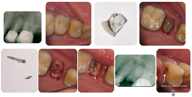
* This photograph shows decay on an adjacent bicuspid. The patient was notified of this lesion. It is now up to the patient to address this issue. Avoidance will obviously result in more destruction of this tooth with the possibility of the need for endodontic therapy and a more expensive final restoration.
** This is providing you are knowlegeable in regards to proper techniques. This involves study and consultation with more experienced practitioners. It also involves your ability to THINK about what you are trying to do. Thomas Watson, the founder of IBM, placed THINK cards in front of all his employees. And, in regards to endodontic therapy, the cleaner the canal, the better the prognosis. I remember once speaking to a professor of endodontics regarding obturation of the canal. He said, it’s not what you put into the canal, it’s what you take out of the canal that’s more important.
PATIENT REPORT
Saturday, September 1, 2012.
Patient presents with pain and facial swelling. Clinical and radiographic exam shows the upper left second bicuspid (tooth # 25, international numbering system) sensitive to percussion.
The radiograph shows a peri-apical radiolucency on this endodontically treated tooth. There appears to be a fractured metal segment within the lingual canal. Treatment options were discussed ( extraction, peri-apical surgery, removal of existing crown, posts and gutta percha in order to establish drainage ). Martin elected to have the third option performed.
Martin was given a Vicodin ES analgesic and a starter dose of antibiotic ( amoxicillin, two 500 mg. tablets ). The porcelain crown/metal crown was removed. Two metal posts were removed along with the fractured metal segment. The remaining gutta percha was removed and drainage was established. The remaining root was left open for continued drainage.
Definitive therapy should include reevaluation of the restorative prognosis (appears favorable if proper endodontics is performed).
Please note that Martin has decay on the adjacent first bicuspid. The periodontal tissues appear inflamed and fibrotic with areas of bleeding on probing. A complete dental examination would be appropriate.