Fixing a deteriorated dentition

(Click on photos to enlarge.)

Initial photo of generalized dentaldeteriorarion
Initial photos show generalized deterioration. As an architect would study the requirements for the restoration of a deteriorated building a dentist basically does the same thing. Important aspects of therapy are not only based on the patient’s desires but also the budget and what the patient wants as compared to what is considered proper therapy .
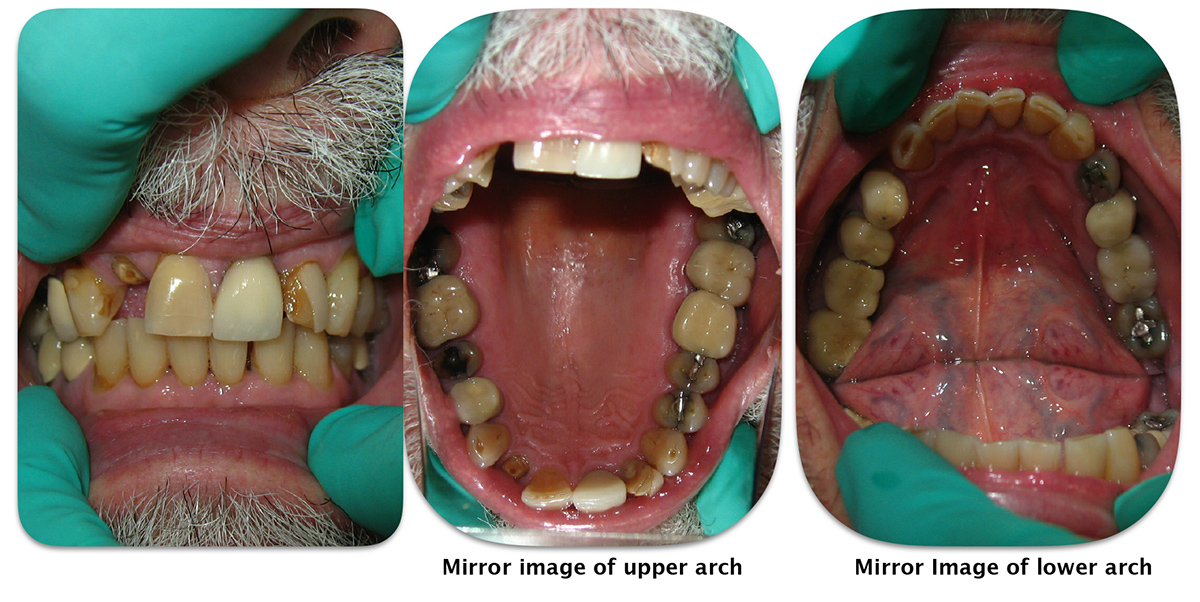
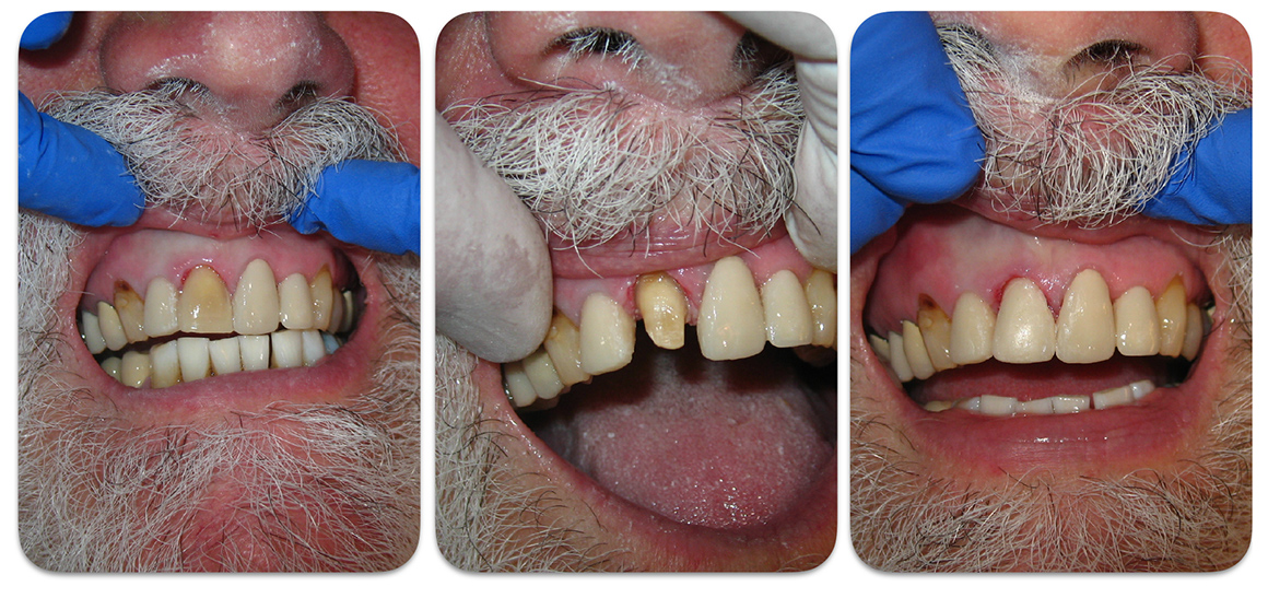
Once treatment alternatives are devised and presented to the patient the therapy can be initiated.
Patient wants upper left lateral incisor restored.
In terms of priorities this handsome patient wanted the upper left lateral incisor restored first.
Radio graphs of upper right lateral incisor.
The radiograph of this upper right lateral incisor shows a calcified canal. As a general dentist who does perform endodontic therapy I elected to refer this patient to the endodontist next door to my office. The reason for this has to do with clinical judgement and maintaining a good rapport with the patient. It’s not a great idea to initiate major therapy with a rather difficult procedure.
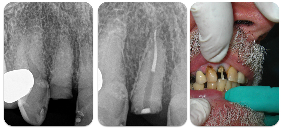
Once the endodontics was completed a carbon fiber post/core was inserted .
Temporary crown fabrication.
A temporary crown was fabricated. Obviously the choice of shade at this point was not a definitive selection.
Asymptomatic upper right second bicuspid restoration.
This asymptomatic upper right second bicuspid was restored via the placement of titanium pins and a composite core.
It is important when placing these pins to utilize high powered magnification. Improper placement can result in perforation of the root or the pulp chamber. This is not an easy procedure. The other option is to perform endodontic therapy so that a more substantial post/core can be fabricated. Of course conservative therapy is many times the best option.
Initial preparations of the upper left central incisor
Initial preparations of the upper left central incisor and the adjacent lateral incisor along with fabrication of temporary crowns.*
* Please note that when performing therapy on vital teeth and soft tissues we utilize septocaine (articaine hydrochloride 4 % and epinephrine 1:100,000). This anesthetic is amazing. Prior to my finding septocaine, I needed to administer mandibular block injections when working on the lower arch. Block injections are still sometimes necessary on occasion. When more hemostasis is required I utilize xylocaine with 1:50,000 epinephrine.
upper right central incisor is prepared for a crown
The upper right central incisor is prepared for a crown and the temporary restoration is fabricated.
Bleaching of the lower teeth
Bleaching of the lower teeth is accomplished with a custom made bleaching tray and the Opalescence system ( 20 % ).
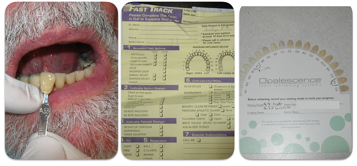
It’s important to utilize a good fitting tray that keeps the bleaching solution adjacent to the crowns and not spilling over the gingival tissues.
Dental improvement after the bleaching
Dramatic improvement after the bleaching.
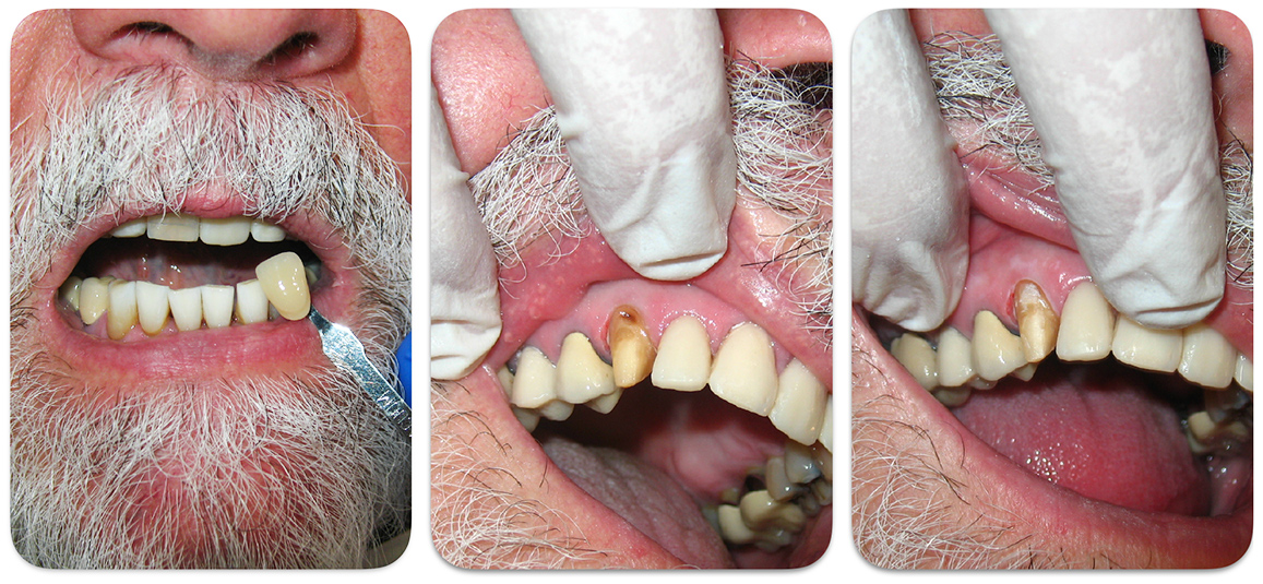
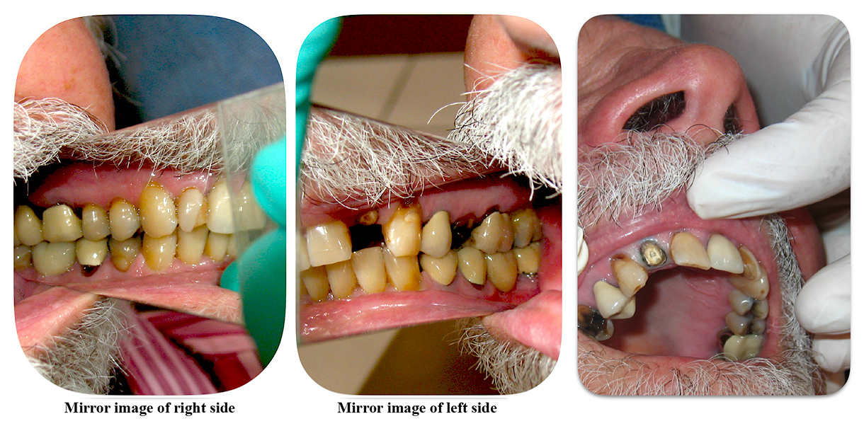
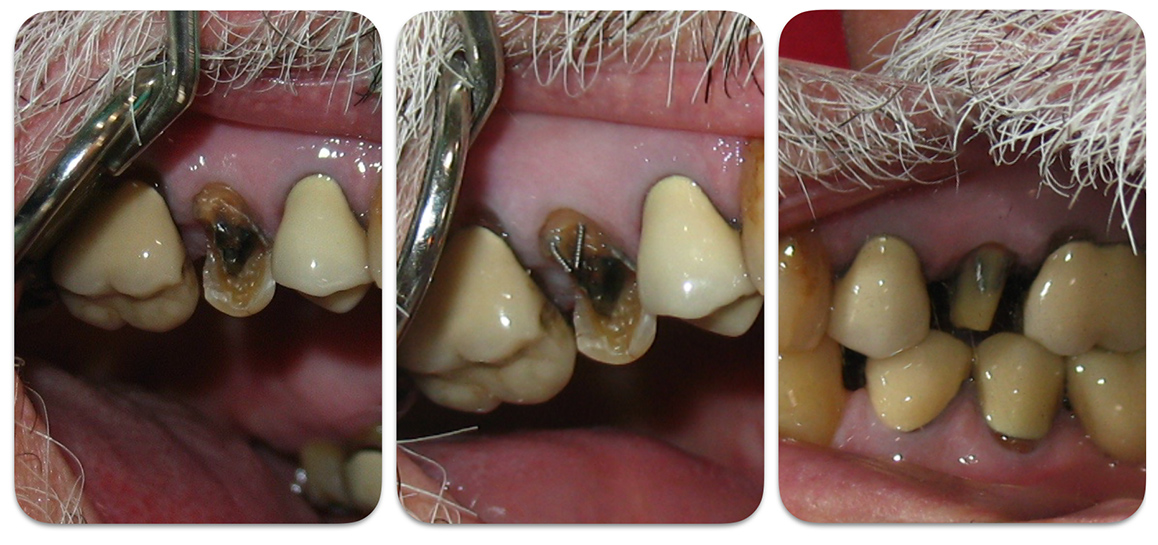
Crown preparation on the upper right cuspid.
Final crowns and a porcelain veneer
The temporary crowns were removed gingival, retraction cord was carefully placed and a final impression was taken. Final crowns were fabricated and a porcelain veneer was made for the upper left cuspid.
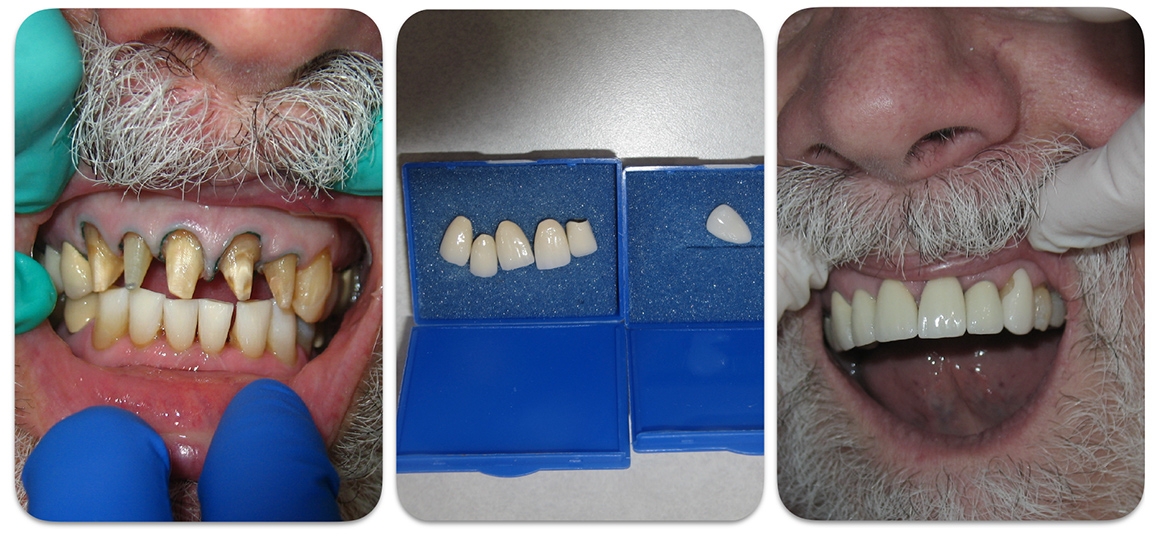
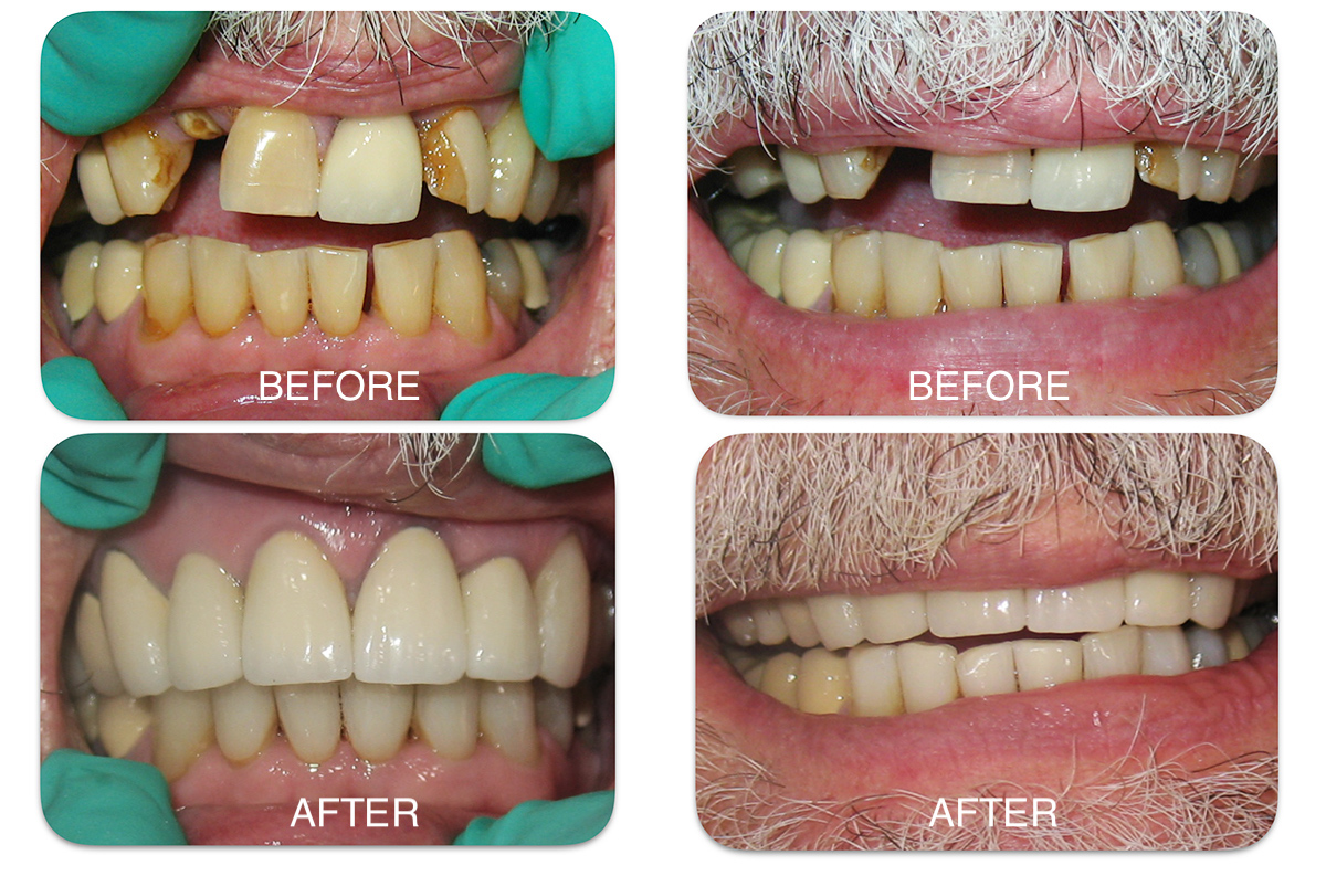
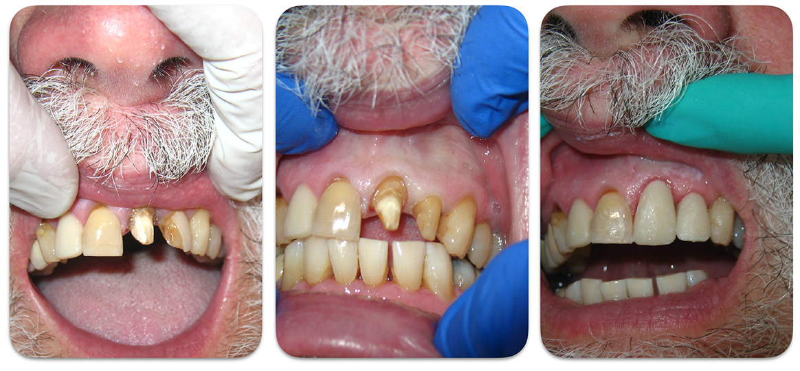
The crowns were initially cemented with temporary cement mixed with a small amount of vaseline so that they are stable when evaluating the occlusion and aesthetics.
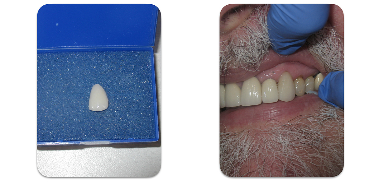
Please note the small porcelain fracture on the cuspid veneer. The prep was modified and a new impression was taken.
new porcelain veneer cemented to the upper left cuspid
The new porcelain veneer is cemented to the upper left cuspid. We utilized 3M ESPE RelyX Unicem 2 cement.
The patient was very pleased with the final result.

Comments are closed.